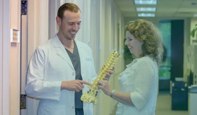
Spondylolisthesis is a condition where one vertebra slips or moves relative to another vertebra.
This term is most often used in the lumbar spine and usually refers to a vertebral body slipping forward on another vertebral body. This malalignment can occur with trauma, a developmental defect in the bone, or, most often, as a degenerative process over many years.
Spondylolisthesis is graded 1-5, with 5 being the most severe and 1 being the most common. Sometimes, the term spondylolysis is used, and this refers to a pars defect, which is typically due to a congenital weakness in the connection between the joints and the spine. This condition may appear in adolescents and may cause progressive back pain, especially with movement throughout one’s life.
With this condition, rarely do patients experience radiating pain and weakness in their lower extremities, and the condition is often found when adolescents engage in contact sports.
In contrast, degenerative spondylolisthesis often affects older individuals and can result in pain, weakness, and numbness in the lower extremities especially with prolonged standing and walking.
Back pain also can occur with degenerative spondylolisthesis. In any of these cases, the problem with spondylolisthesis is vertebrae slippage, and early detection of this spinal condition can reduce symptom severity and quicken the time to recovery.
Spondylolisthesis often manifests as back pain. Mechanical back pain is back pain that occurs with movement and stops or improves significantly when movement stops.
Mechanical back pain occurs more with dynamic spondylolisthesis—that is, vertebral slippage that becomes worse and better with flexion and extension of the spine, respectively.
With degenerative spondylolisthesis, additional symptoms are lower extremity pain, weakness, tingling, burning, and numbness. These symptoms often are put under the umbrella of sciatica and are the result of nerve compression.
Depending on the severity and grade of spondylolisthesis, the associated symptoms can be transient or constant, but early medical evaluation and diagnosis are paramount if any of these symptoms are present and persist.
While spondylolisthesis can be suspected based on clinical examination and symptoms, CT scans and X-rays can be used to confirm the diagnosis. Often, upright X-rays are done with flexion and extension views to determine if there is vertebral slippage and if it is dynamic or static in nature.
Additional spinal imaging includes an MRI, which can verify if the neural elements are compressed along with the spondylolisthesis diagnosis. The grade of spondylolisthesis also can be measured with any of these imaging modalities.
It is important to have both an accurate diagnosis of spondylolisthesis and understand the source of symptoms (whether it is nerve compression and/or bony movement) to tailor specific treatments to individual patients.
There are many non-surgical ways to treat spondylolisthesis.
Physical therapy is useful to strengthen the muscles of the spine and core in an attempt to reduce vertebral slippage.
Spinal bracing can also reduce slippage and can be a trial to see if symptoms would be reduced with fusion surgery.
Pain management, including oral medications and pain-directed injections, can be helpful to calm symptoms in milder cases of spondylolisthesis and may reduce symptoms if surgery is being planned.
Lifestyle changes, including weight loss, avoiding contact sports, and avoiding heavy-duty jobs, can improve symptoms. If any of these non-surgical spondylolisthesis treatments fail to provide sufficient relief or if lifestyle modifications like changing jobs and avoiding sports are too cumbersome for patients, it may be time to consider surgical treatment for lasting relief.
The most common spondylolisthesis surgery involves fusing the vertebrae, which causes the bones to grow together into one bone. During this process, the vertebral bodies are held in place using devices like rods and screws.
At Performance Spine and Brain, our neurosurgeon, Dr. Birinyi, performs most of these spinal fusion surgeries in a minimally invasive fashion.
These techniques involve making small incisions and minimizing the disruption of the spinal musculature to reduce procedural blood loss, minimize postoperative pain, and hasten a return to normal life.
In some instances, the surgeries can be performed in the outpatient setting. In addition, in cases with individuals who may not be able to tolerate a fusion surgery or may not want to undergo such a procedure, if spondylolisthesis is static in nature, Dr. Birinyi can utilize minimally invasive decompressive techniques and avoid a fusion and spinal instrumentation.
In any case, surgery becomes necessary when non-operative therapies fail, and patients find that their symptoms are extremely disruptive to their daily lives.
When surgery is performed, a bony fusion often takes place in the lumbar spine within six to twelve months, and there will be some activity limitations for patients during this time.
Many patients are able to return to reasonable daily activities, though much sooner, in a matter of a few months. With a simple decompression without a fusion, patients often return to reasonably normal activities within a few weeks.
The recovery from spondylolisthesis surgery varies significantly based on many variables, including age, health status, preoperative pain, and surgical techniques used. Typically, patients undergoing surgery with minimally invasive techniques have less postoperative pain compared to those undergoing surgery with open techniques and thus leave the hospital sooner.
Following any surgery, nursing staff and therapists work with patients to ensure that they can safely care for themselves. Sometimes, an inpatient physical rehabilitation stay is necessary for a matter of days or weeks.
During the process of healing from a lumbar spinal fusion, it is important for patients not to bend and twist in their lumbar spine, and lifting should be minimized. These lifting and movement restrictions will be relaxed gradually over several months until the surgery recovery has been completed.
Individuals with desk jobs often can return to work within two to four weeks after a fusion surgery. Individuals with high-intensity and physically demanding jobs may not be able to return to full-duty work for six to twelve months.
Wearing a lumbar brace following surgery is beneficial for restricting movement and preventing possible damage to surgical hardware. Bone growth stimulators can be used to hasten the fusion process as an added measure to ensure a successful surgical recovery.
Physical therapy can begin in the water a month after surgery, whereas land-based physical therapy often starts three months following surgery.
The key is to heal appropriately the first time and abide by restrictions very closely, focusing on the long-term goal of a healed and successful spine surgery with a great long-term outlook and reduced or no symptoms.
